
Exams are done at our Lincoln Nebraska Location
What are diabetes eye exams?
Routine eye examinations can reveal a lot of different things about a patient like what type of prescription grade the glasses they need or what kind of contact lenses would be best for them. On the other hand, diabetic eye exams are specifically done to detect whether or not a patient has diabetes. What a patient has diabetes, the eyes are usually implicated and could incurred considerable damage as the condition progresses. Usually, the smaller blood vessels in the patient’s retina become affected and this results in a condition called diabetic retinopathy. Regular eye examinations are very important in the early detection of diabetic retinopathy because the signs will only be discovered under the close inspection of a diabetic eye exam. This means that the patient may not even know that their eyes are getting damaged in the process.
How often do I need to get a diabetic eye exam?
It is recommended that a patient go in for a regular eye check once every year or once into years by the doctor that is qualified to carry out a diabetic eye exam. If the patient’s diabetes is already in its advanced stages, then he may need to see the eye doctor for his diabetic eye exams more often.
And what types of diabetic eye exam are there?
- A visual actually test maybe administered as part of a diabetic eye exam to measure the patient’s ability to focus on objects at different distances. This test allows doctors to detect if there is any degree of vision loss or other eye problems that the patient should be concerned about.
- A gonioscopy test is used to determine the drainage angle of the eye and further test if the fluids inside the eye are being drained properly. This is essential for patients also suspected of having glaucoma
- Tonometry is a test that may be administered during a diabetic eye exam to measure the intraocular pressure of the eye in an effort to detect the presence of glaucoma. It has been found that having diabetes increases the patient’s risk of developing this disease as well.
- Ophthalmoscopy and slit lamp exams are tests that the doctor uses to examine the structures within the eye to possibly detect the presence of cataracts or to look for abnormalities within the retina.
- An optical coherence tomography is a test that checks for fluid retention within the retina
 What happens during a diabetic eye exam?

- A diabetic eye exam is similar to a regular eye exam in the sense that the doctor has to check the patient’s vision using a Snellen chart made up of random letters and arranged in different sizes.
- The doctor will administer eye drops directly into the eyes and this will help him have a better view of the back of the eye. These eye drops are especially formulated to dilate the pupils of the eyes, but the patient may feel a singing sensation when the drops are administered initially. Some patients have also reported a metallic taste in the mouth during this step.
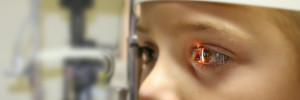
- Using a machine that can both magnify and illuminate the eyes, the doctor can begin to assess certain areas of the eyes such as the blood vessels from the front to the middle section of the eyes, the back portion of the eye, and the optic nerve itself. Being able to look into these areas will allow doctors to check for any sign of damage caused being by diabetes.
- Another part of the diabetic eye exam is the examination of the surface of the patient’s eye for clarity using a slit lamp
- During the course of a diabetic eye exam, doctors may take photos of that area’s of the eyes being viewed so that a more detailed inspection can be done in addition to the actual eye examination.
Are there any risks to getting a diabetic eye exam?
The risk involved with a diabetic eye exam usually comes with the administration of the eye drops that are used to dilate the pupils of the eyes during the examination. Some patients have reported that they experienced blurry vision up to six hours after the administration of the dilating solution. Because of this, it will also be harder for the patient to focus on the objects that they are looking at until the effects of the solution has worn off. In relation to this, patients must take care to protect their eyes from the direct glare of the sun or other light sources because the dilated pupils will make the eyes highly sensitive to light for as long as the effects of the solution are active.